


Childbirth About nine months after fertilization, the fetus is ready for birth. A complex set of factors triggers the process; one of these factors is the release of the hormone oxytocin from the mother's posterior pituitary gland. Oxytocin affects a group of large involuntary muscles in the uterine wall. As these muscles are stimulated, they begin a series of rhythmic contractions collectively known as labor. As labor progresses, the contractions become more frequent and more powerful. The opening of the cervix expands until it is large enough for the head of the baby to pass through. At some point, the amniotic sac breaks, and the fluid it contains rushes out of the vagina. Contractions of the uterus force the baby, usually head first, out through the vagina.
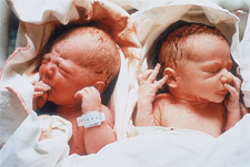
FIGURE 34–24 Newborns Twins, ten minutes after birth, adjusting to life outside the uterus.
As the baby meets the outside world, he or she may begin to cough or cry, a process that rids the lungs of fluid. Breathing starts almost immediately, and the blood supply to the placenta begins to dry up. The umbilical cord is clamped and cut, leaving a small piece attached to the baby. This piece will soon dry and fall off, leaving a scar known as the navel—or, its more familiar term, the belly button. In a final series of uterine contractions, the placenta itself and the now-empty amniotic sac are expelled from the uterus as the afterbirth.
The baby now begins an independent existence. Most newborns are remarkably hardy. Their systems quickly make the switch to life outside the uterus, supplying their own oxygen, excreting wastes on their own, and maintaining their own body temperatures.
The interaction of the mother's reproductive and endocrine systems does not end at childbirth. Within a few hours after birth, the pituitary hormone prolactin stimulates the production of milk in the breast tissues of the mother. The nutrients present in that milk contain everything the baby needs for growth and development during the first few months of life.
Quick Lab
GUIDED INQUIRY
Embryonic Development
Use a dropper pipette to transfer several early-stage frog embryos in water to a depression slide. CAUTION: Handle glass slides with care.
Look at the embryos under the dissecting microscope at low power. Sketch what you see.
Look at the prepared slides of the early embryonic stages of a frog. Make sketches of what you see.
Observe Describe any differences you saw among the cells. At what stage is cell differentiation visible?
Observe Were you able to see a distinct body plan? At what stage did the body plan become visible?
Draw Conclusions Describe any organs you saw. At what stage did specific organs form?
Analyze and Conclude
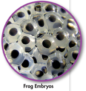
Table of Contents
- Formulas and Equations
- Applying Formulas and Equations
- Mean, Median, and Mode
- Estimation
- Using Measurements in Calculations
- Effects of Measurement Errors
- Accuracy
- Precision
- Comparing Accuracy and Precision
- Significant Figures
- Calculating With Significant Figures
- Scientific Notation
- Calculating With Scientific Notation
- Dimensional Analysis
- Applying Dimensional Analysis