


Technology & Biology
Testing for Heart Disease
Ever-improving imaging techniques make it possible for doctors to diagnose heart disease and disorders quickly and without the risk of invasive procedures. None of these tests involves inserting instruments into the body, but they reveal the inner workings of the heart with remarkable accuracy.
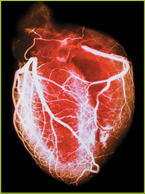
Computed Tomography Angiography
A patient is injected with an iodine-based dye. Then the CT scanner rotates over the patient and takes multiple X-rays of the heart, which a computer uses to form three-dimensional images. The test can show if parts of blood vessels are blocked or damaged. The results can be used to determine what further tests are needed or as a guide for planning surgery.
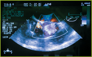
Echocardiography
High-frequency sound waves, transmitted through the chest, are fed into a computer, which analyzes the “echoes” to produce moving images of the heart. This is an especially safe test because it doesn't involve radiation or dyes. The test allows doctors to see the heart in action. It can reveal an enlarged heart, reduced pumping action, and structural problems.
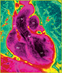
Magnetic Resonance Imaging (MRI)
MRI uses powerful magnets to produce images that are particularly good for examining muscle and other soft tissue. Professionals analyzing MRI images can see the difference between healthy tissue and unhealthy tissue. MRI does not involve radiation or iodine-based dyes. It can be used to assess heart muscle damage caused by a heart attack, birth defects, or abnormal growths.
WRITING
In a paragraph, explain which technique would most likely be used to check for advanced atherosclerosis in a coronary artery.
Table of Contents
- Formulas and Equations
- Applying Formulas and Equations
- Mean, Median, and Mode
- Estimation
- Using Measurements in Calculations
- Effects of Measurement Errors
- Accuracy
- Precision
- Comparing Accuracy and Precision
- Significant Figures
- Calculating With Significant Figures
- Scientific Notation
- Calculating With Scientific Notation
- Dimensional Analysis
- Applying Dimensional Analysis