


32 Assessment
32.1 The Skeletal System
Understand Key Concepts
The network of tubes that runs through compact bone is called the
periosteum.
joint.
Haversian canals.
marrow.
What occurs during ossification?
Bones lose minerals and mass.
Cartilage is replaced by bone.
Vitamin
is synthesized.
Bones fracture more easily.
Small sacs of synovial fluid that help reduce friction between the bones of a joint are called
bursae.
ligaments.
tendons.
cartilage.
What types of tissues are found in the skeletal system?
What is the advantage of spongy bone tissue in the ends of long bones?
Draw a diagram of a long bone and label the structures.
Which type of freely movable joint allows for the most range of motion?
Think Critically
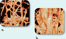
Interpret Visuals Which bone sample shows signs of osteoporosis, choice a or choice b? Explain.
Infer Disks of rubbery cartilage are found between the individual bones in the spinal column. What function do you think these disks serve?
Predict Blood vessels bring oxygen and nutrients to all parts of the body. Ligaments contain fewer blood vessels than some other kinds of tissues contain. How might this situation affect the rate of healing in injured ligaments? Explain.
Use Models Suppose you want to build a robotic arm that works the way the human elbow works. Describe or sketch three facts about the elbow that you could use in your planning.
32.2 The Muscular System
Understand Key Concepts
In which part of the body would you find striated muscle tissue with relatively small cells that have one or two nuclei?
thigh
stomach
blood vessels
heart
Two proteins that are involved in the contraction of muscle are
sarcomere and myofibril.
actin and myosin.
periosteum and cartilage.
ATP and acetylcholine.
The point of contact between a motor neuron and a skeletal muscle cell is called a
cross-bridge site.
gap junction.
sarcomere.
neuromuscular junction.
Describe the primary function of each of the three types of muscle tissue.
Use the sliding-filament model to describe how skeletal muscles work.
Describe how the release of acetylcholine from a motor neuron affects a muscle cell.
Explain this statement: “Most skeletal muscles work in opposing pairs.”
What is one difference between the structure of fast-twitch and slow-twitch muscle fibers? How does this difference relate to their functions?
Table of Contents
- Formulas and Equations
- Applying Formulas and Equations
- Mean, Median, and Mode
- Estimation
- Using Measurements in Calculations
- Effects of Measurement Errors
- Accuracy
- Precision
- Comparing Accuracy and Precision
- Significant Figures
- Calculating With Significant Figures
- Scientific Notation
- Calculating With Scientific Notation
- Dimensional Analysis
- Applying Dimensional Analysis