


7.1 Life Is Cellular
 What is the cell theory?
- How do microscopes work?
- How are prokaryotic and eukaryotic cells different?
cell • cell theory • cell membrane • nucleus • eukaryote • prokaryote
Outline Before you read, make an outline using the green and blue headings in the text. As you read, fill in notes under each heading.
THINK ABOUT IT What's the smallest part of any living thing that still counts as being “alive”? Is a leaf alive? How about your big toe? How about a drop of blood? Can we just keep dividing living things into smaller and smaller parts, or is there a point at which what's left is no longer alive? As you will see, there is such a limit, the smallest living unit of any organism—the cell.
The Discovery of the Cell
What is the cell theory?
“Seeing is believing,” an old saying goes. It would be hard to find a better example of this than the discovery of the cell. Without the instruments to make them visible, cells remained out of sight and, therefore, out of mind for most of human history. All of this changed with a dramatic advance in technology—the invention of the microscope.
Early Microscopes In the late 1500s, eyeglass makers in Europe discovered that using several glass lenses in combination could magnify even the smallest objects to make them easy to see. Before long, they had built the first true microscopes from these lenses, opening the door to the study of biology as we know it today.
In 1665, Englishman Robert Hooke used an early compound microscope to look at a nonliving thin slice of cork, a plant material. Under the microscope, cork seemed to be made of thousands of tiny empty chambers. Hooke called these chambers “cells” because they reminded him of a monastery's tiny rooms, which were called cells. The term cell is used in biology to this day. Today we know that living cells are not empty chambers, that in fact they contain a huge array of working parts, each with its own function.
In Holland around the same time, Anton van Leeuwenhoek used a single-lens microscope to observe pond water and other things. To his amazement, the microscope revealed a fantastic world of tiny living organisms that seemed to be everywhere, in the water he and his neighbors drank, and even in his own mouth. Leeuwenhoek's illustrations of the organisms he found in the human mouth—which today we call bacteria—are shown in Figure 7–1.
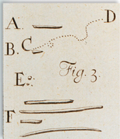
FIGURE 7–1 Early Microscope Images Using a simple microscope, Anton van Leeuwenhoek was the first to observe living microorganisms. These drawings, taken from one of his letters, show bacteria in the human mouth.

Table of Contents
- Formulas and Equations
- Applying Formulas and Equations
- Mean, Median, and Mode
- Estimation
- Using Measurements in Calculations
- Effects of Measurement Errors
- Accuracy
- Precision
- Comparing Accuracy and Precision
- Significant Figures
- Calculating With Significant Figures
- Scientific Notation
- Calculating With Scientific Notation
- Dimensional Analysis
- Applying Dimensional Analysis