


Technology & Biology
Studying the Brain and Addiction
Studies at the National Institute of Drug Abuse (NIDA) have demonstrated why drugs that stimulate dopamine produce a pattern of addiction that is difficult to break. The brain is a flexible organ that responds to its environment and continually adjusts its internal chemistry. When it senses increased levels of dopamine, it adjusts by cutting down on the number of receptors for the neurotransmitter.
NIDA researchers used a powerful imaging technique known as positron emission tomography (PET) to visualize the density of dopamine receptors in brains affected by drug addition, and the results, shown here, are striking. Brains of individuals abusing alcohol and illegal drugs show dramatically lower concentrations of dopamine receptors than the brains of individuals not abusing the drugs.
Positron emission tomography (PET) allows researchers to visualize labeled molecules deep inside the body. PET is routinely used to pinpoint regions of cellular activity. To locate dopamine receptors, a molecule that binds to the receptor is labeled with a radioactive isotope of carbon. Within a few minutes, the isotope emits a subatomic particle called a positron. The location of the particle is revealed by gamma rays released when it collides with other particles. By locating thousands of positron emissions, computers can put together detailed images showing the location of the labeled molecules.
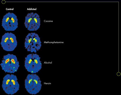
In these images, areas of highest dopamine receptor density appear red. Areas of lowest dopamine receptor density appear green.
WRITING
Using the information in this feature, create a poster to discourage peers from using addictive drugs.
Table of Contents
- Formulas and Equations
- Applying Formulas and Equations
- Mean, Median, and Mode
- Estimation
- Using Measurements in Calculations
- Effects of Measurement Errors
- Accuracy
- Precision
- Comparing Accuracy and Precision
- Significant Figures
- Calculating With Significant Figures
- Scientific Notation
- Calculating With Scientific Notation
- Dimensional Analysis
- Applying Dimensional Analysis