


GROUPS OF BACTERIA
There is no generally agreed phylogeny for the bacteria. Included here are some of the major groups within the domain.
Helicobacter pylori is rod-shaped and has several flagella used for movement. This bacterium infects the stomach lining and causes ulcers in some people. (TEM 7100X)
PROTEOBACTERIA
This large and diverse clade of bacteria includes Escherichia (E. coli), Salmonella, Helicobacter, and the nitrogen-fixing soil bacterium Rhizobium.

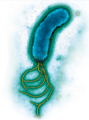
The spiral-shaped bacterium that causes syphilis is Treponema pallidum. (SEM 10,000X)
SPIROCHAETES
The spirochaetes (SPY roh keets) are named for their distinctive spiral shape. They move in a corkscrew-like fashion, twisting along as they are propelled by flagella on both ends of the cell. Most are free-living, but a few cause serious diseases, including syphilis, Lyme disease, and leptospirosis.
ACTINOBACTERIA
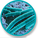
A large number of soil bacteria belong to this group. Some form long filaments. Members include the Streptomyces and Actinomyces, which are natural producers of many antibiotics, including streptomycin. A related group is the Firmicutes. The Firmicutes include Bacillus anthracis (anthrax), Clostridia (tetanus and botulism), and Bacillus thuringensis, which produces a powerful insecticide used for genetic engineering in plants.
Chains of spores of soil bacteria, genus Streptomyces (SEM 3400X)
CYANOBACTERIA
The cyanobacteria are photosynthetic prokaryotes that were once called “blue-green algae.” They are among the oldest organisms on Earth, having been identified in rocks dating to more than 3 billion years ago. They are found in salt water and fresh water, in the soil, and even on the surfaces of damp rocks. They are the only organisms on Earth that are able to fix carbon and nitrogen under aerobic conditions, and this enables them to play critical roles in the global ecosystem, where they serve as key sources of carbon and nitrogen.
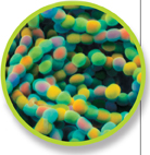
Many cyanobacteria form long filaments of attached cells, like those shown here (genus Lyngbya, SEM 540X).
• A Closer Look
The Gram Stain
A Microbiologist's Quick Diagnostic
The Gram stain, developed by the nineteenth-century Danish physician Hans Christian Gram, allows microbiologists to categorize bacteria quickly into one of two groups based on their cell wall composition. Gram-positive bacteria lack a membrane outside the cell wall and take up the stain easily. Gram-negative bacteria, on the other hand, have an outer membrane of lipids and carbohydrates that prevents them from absorbing the gram stain. Many gram-negative bacteria are found among the proteobacteria. On the other hand, actinobacteria are mostly gram-positive.
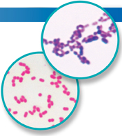
Gram-positive bacteria appear purple after staining, while gram-negative bacteria appear pink. (LM 1000X)
Table of Contents
- Formulas and Equations
- Applying Formulas and Equations
- Mean, Median, and Mode
- Estimation
- Using Measurements in Calculations
- Effects of Measurement Errors
- Accuracy
- Precision
- Comparing Accuracy and Precision
- Significant Figures
- Calculating With Significant Figures
- Scientific Notation
- Calculating With Scientific Notation
- Dimensional Analysis
- Applying Dimensional Analysis