


Use of the Microscope
The microscope used in most biology classes, the compound microscope, contains a combination of lenses. The eyepiece lens is located in the top portion of the microscope. This lens usually has a magnification of 10X. Other lenses, called objective lenses, are at the bottom of the body tube on the revolving nosepiece. By rotating the nosepiece, you can select the objective through which you will view your specimen.
The shortest objective is a low-power magnifier, usually 10×. The longer ones are of high power, usually up to 40× or 43×. The magnification is marked on the objective. To determine the total magnification, multiply the magnifying power of the eyepiece by the magnifying power of the objective. For example, with a 10× eyepiece and a 40× objective, the total magnification is 10 × 40 = 400X.
Learning the name, function, and location of each of the microscope's parts is necessary for proper use. Use the following procedures when working with the microscope.
Carry the microscope by placing one hand beneath the base and grasping the arm of the microscope with the other hand.
Gently place the microscope on the lab table with the arm facing you. The microscope's base should be resting evenly on the table, approximately 10 cm from the table's edge.
Raise the body tube by turning the coarse adjustment knob until the objective lens is about 2 cm above the opening of the stage.
Rotate the nosepiece so that the low-power objective (10×) is directly in line with the body tube. A click indicates that the lens is in line with the opening of the stage.
Look through the eyepiece and switch on the lamp or adjust the mirror so that a circle of light can be seen. This is the field of view. Moving the lever of the diaphragm permits a greater or smaller amount of light to come through the opening of the stage.
Place a prepared slide on the stage so that the specimen is over the center of the opening. Use the stage clips to hold the slide in place.
Look at the microscope from the side. Carefully turn the coarse adjustment knob to lower the body tube until the low-power objective almost touches the slide or until the body tube can no longer be moved. Do not allow the objective to touch the slide.
Look through the eyepiece and observe the specimen. If the field of view is out of focus, use the coarse adjustment knob to raise the body tube while looking through the eyepiece. CAUTION: To prevent damage to the slide and the objective, do not lower the body tube using the coarse adjustment while looking through the eyepiece. Focus the image as best you can with the coarse adjustment knob. Then, use the fine adjustment knob to focus the image more sharply. Keep both eyes open when viewing a specimen. This helps prevent eyestrain.
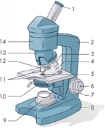
Table of Contents
- Formulas and Equations
- Applying Formulas and Equations
- Mean, Median, and Mode
- Estimation
- Using Measurements in Calculations
- Effects of Measurement Errors
- Accuracy
- Precision
- Comparing Accuracy and Precision
- Significant Figures
- Calculating With Significant Figures
- Scientific Notation
- Calculating With Scientific Notation
- Dimensional Analysis
- Applying Dimensional Analysis