


The Cell Theory Soon after van Leeuwenhoek, observations by scientists made it clear that cells are the basic units of life. In 1838, German botanist Matthias Schleiden concluded that all plants are made of cells. The next year, German biologist Theodor Schwann stated that all animals are made of cells. In 1855, German physician Rudolf Virchow concluded that new cells can be produced only from the division of existing cells, confirming a suggestion made by German Lorenz Oken 50 years earlier. These discoveries, confirmed by many biologists, are summarized in the cell theory, a fundamental concept of biology.  The cell theory states:
All living things are made up of cells.
Cells are the basic units of structure and function in living things.
New cells are produced from existing cells.
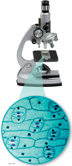
FIGURE 7–2 Light Microscope and Cell Stains This specimen of onion leaf skin has been stained with a compound called toluidine blue. The dye makes the cell boundaries and nuclei clearly visible.
Exploring the Cell
How do microscopes work?
A microscope, as you know, produces an enlarged image of something very small. Most microscopes use lenses to magnify the image of an object by focusing light or electrons. Following in the footsteps of Hooke, Virchow, and others, modern biologists still use microscopes to explore the cell. But today's researchers use technology more powerful than the pioneers of biology could ever have imagined.
Light Microscopes and Cell Stains The type of microscope you are probably most familiar with is the compound light microscope. A typical light microscope allows light to pass through a specimen and uses two lenses to form an image. The first lens, called the objective lens, is located just above the specimen. This lens enlarges the image of the specimen. Most light microscopes have several objective lenses so that the power of magnification can be varied. The second lens, called the ocular lens, magnifies this image still further. Unfortunately, light itself limits the detail, or resolution, of images in a microscope. Like all forms of radiation, lightwaves are diffracted, or scattered, as they pass through matter. Because of this, light microscopes can produce clear images of objects only to a magnification of about 1000 times.
Another problem with light microscopy is that most living cells are nearly transparent. Using chemical stains or dyes, as in Figure 7–2, can usually solve this problem. Some of these stains are so specific that they reveal only certain compounds or structures within the cell. Many of the slides you'll examine in your biology class laboratory will be stained this way.
A powerful variation on these staining techniques uses dyes that give off light of a particular color when viewed under specific wavelengths of light, a property called fluorescence. Fluorescent dyes can be attached to specific molecules and can then be made visible using a special fluorescence microscope. New techniques, in fact, enable scientists to engineer cells that attach fluorescent labels of different colors to specific molecules as they are produced. Fluorescence microscopy makes it possible to see and identify the locations of these molecules and even allows scientists to watch them move around in a living cell.
Table of Contents
- Formulas and Equations
- Applying Formulas and Equations
- Mean, Median, and Mode
- Estimation
- Using Measurements in Calculations
- Effects of Measurement Errors
- Accuracy
- Precision
- Comparing Accuracy and Precision
- Significant Figures
- Calculating With Significant Figures
- Scientific Notation
- Calculating With Scientific Notation
- Dimensional Analysis
- Applying Dimensional Analysis