


Electron Microscopes Light microscopes can be used to see cells and cell structures as small as 1 millionth of a meter—certainly pretty small! But what if scientists want to study something smaller than that, such as a virus or a DNA molecule? For that, they need electron microscopes. Instead of using light, electron microscopes use beams of electrons that are focused by magnetic fields. Electron microscopes offer much higher resolution than light microscopes. Some types of electron microscopes can be used to study cellular structures that are 1 billionth of a meter in size.
There are two major types of electron microscopes: transmission and scanning. Transmission electron microscopes make it possible to explore cell structures and large protein molecules. But because beams of electrons can only pass through thin samples, cells and tissues must be cut into ultrathin slices before they can be examined. This is the reason that such images often appear flat and two dimensional.
In scanning electron microscopes, a pencil-like beam of electrons is scanned over the surface of a specimen. Because the image is formed at the specimen's surface, samples do not have to be cut into thin slices to be seen. The scanning electron microscope produces stunning three-dimensional images of the specimen's surface.
Electrons are easily scattered by molecules in the air, which means samples must be placed in a vacuum to be studied with an electron microscope. As a result, researchers must chemically preserve their samples. Electron microscopy, then, can only be used to examine nonliving cells and tissues.
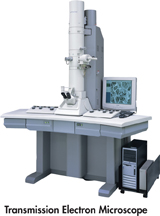
FIGURE 7–3 Micrographs Different types of microscopes can be used to examine cells. Here, yeast cells are shown in a light micrograph (LM 500X), transmission electron micrograph (TEM 4375X), and a scanning electron micrograph (SEM 3750X).
Infer If scientists were studying a structure found on the surface of yeast, which kind of microscope would they likely use?
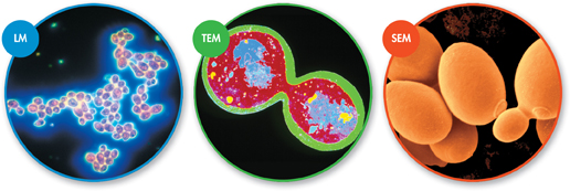
Look at Figure 7–3, which shows yeast cells as they might look under a light microscope, transmission electron microscope, and scanning electron microscope. You may wonder why the cells appear to be different colors in each micrograph. (A micrograph is a photo of an object seen through a microscope.) The colors in light micrographs come from the cells themselves, or from the stains and dyes used to highlight them. Electron micrographs, however, are actually black and white. Electrons, unlike light, don't come in colors. So scientists often use computer techniques to add “false color” to make certain structures stand out.
 In Your Notebook You are presented with a specimen to examine. What are two questions you would ask to determine the best microscope to use?
Table of Contents
- Formulas and Equations
- Applying Formulas and Equations
- Mean, Median, and Mode
- Estimation
- Using Measurements in Calculations
- Effects of Measurement Errors
- Accuracy
- Precision
- Comparing Accuracy and Precision
- Significant Figures
- Calculating With Significant Figures
- Scientific Notation
- Calculating With Scientific Notation
- Dimensional Analysis
- Applying Dimensional Analysis