


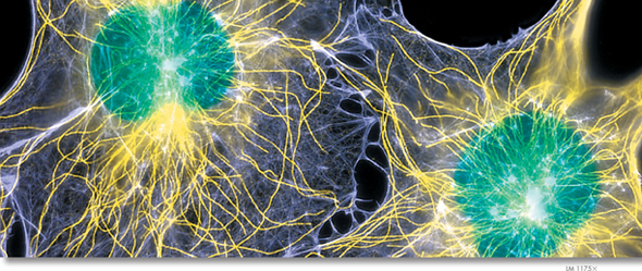
FIGURE 7–9 Cytoskeleton The cytoskeleton supports and gives shape to the cell, and is involved in many forms of cell movement. These connective tissue fibroblast cells have been treated with fluorescent tags that bind to certain elements. Microfilaments are pale purple, microtubules are yellow, and the nuclei are green.
The Cytoskeleton As you know, a factory building is supported by steel or cement beams and by columns that hold up its walls and roof. Eukaryotic cells are given their shape and internal organization by a network of protein filaments known as the cytoskeleton. Certain parts of the cytoskeleton also help transport materials between different parts of the cell, much like the conveyor belts that carry materials from one part of a factory to another. Cytoskeletal components may also be involved in moving the entire cell as in cell flagella and cilia.  The cytoskeleton helps the cell maintain its shape and is also involved in movement. Fluorescence imaging, as seen in Figure 7–9, clearly shows the complexity of a cell's cytoskeletal network. Microfilaments (pale purple) and microtubules (yellow) are two of the principal protein filaments that make up the cytoskeleton.
▸ Microfilaments Microfilaments are threadlike structures made up of a protein called actin. They form extensive networks in some cells and produce a tough flexible framework that supports the cell. Microfilaments also help cells move. Microfilament assembly and disassembly are responsible for the cytoplasmic movements that allow amoebas and other cells to crawl along surfaces.
▸ Microtubules Microtubules are hollow structures made up of proteins known as tubulins. In many cells, they play critical roles in maintaining cell shape. Microtubules are also important in cell division, where they form a structure known as the mitotic spindle, which helps to separate chromosomes. In animal cells, organelles called centrioles are also formed from tubulins. Centrioles are located near the nucleus and help organize cell division. Centrioles are not found in plant cells.
Microtubules also help build projections from the cell surface—known as cilia (singular: cilium) and flagella (singular: flagellum)—that enable cells to swim rapidly through liquid. The microtubules in cilia and flagella are arranged in a “9 + 2” pattern, as shown in Figure 7–10. Small cross-bridges between the microtubules in these organelles use chemical energy to pull on, or slide along, the microtubles, producing controlled movements.
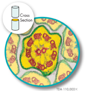
FIGURE 7–10 The “9 + 2” Pattern of Microtubules In this micrograph showing the cross section of a cilium, you can clearly see the 9 + 2 arrangement of the red microtubules. Apply Concepts What is the function of cilia?
Table of Contents
- Formulas and Equations
- Applying Formulas and Equations
- Mean, Median, and Mode
- Estimation
- Using Measurements in Calculations
- Effects of Measurement Errors
- Accuracy
- Precision
- Comparing Accuracy and Precision
- Significant Figures
- Calculating With Significant Figures
- Scientific Notation
- Calculating With Scientific Notation
- Dimensional Analysis
- Applying Dimensional Analysis