


VISUAL SUMMARY
CLUES TO THE STRUCTURE OF DNA
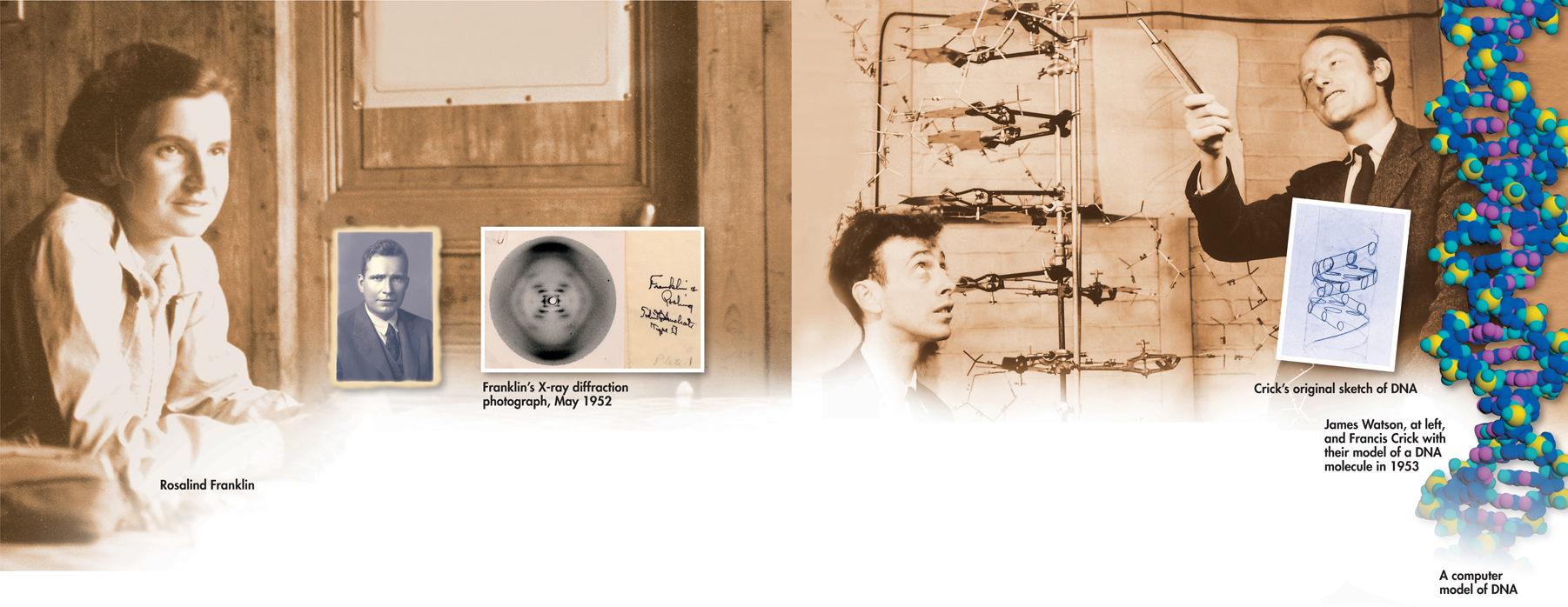
FIGURE 12–6 Erwin Chargaff, Rosalind Franklin, James Watson, and Francis Crick were among the many scientists who helped solve the puzzle of DNA's molecular structure. Franklin's X-ray diffraction photograph shows the pattern that indicated the structure of DNA is helical.
BUILD Vocabulary
ACADEMIC WORDS In biochemistry, the noun helix refers to an extended spiral chain of units in a protein, nucleic acid, or other large molecule. The plural term is helices.
Franklin's X-Rays In the early 1950s, the British scientist Rosalind Franklin began to study DNA. Franklin used a technique called X-ray diffraction to get information about the structure of the DNA molecule. First, she purified a large amount of DNA, then stretched the DNA fibers in a thin glass tube so that most of the strands were parallel. Next, she aimed a powerful X-ray beam at the concentrated DNA samples and recorded the scattering pattern of the X-rays on film. Franklin worked hard to obtain better and better patterns from DNA until the patterns became clear. The result of her work is the X-ray photograph shown in Figure 12–6, taken in the summer of 1952.
By itself, Franklin's X-ray pattern does not reveal the structure of DNA, but it does carry some very important clues. The X-shaped pattern shows that the strands in DNA are twisted around each other like the coils of a spring, a shape known as a helix. The angle of the X suggests that there are two strands in the structure. Other clues suggest that the nitrogenous bases are near the center of the DNA molecule.
The Work of Watson and Crick While Franklin was continuing her research, James Watson, an American biologist, and Francis Crick, a British physicist, were also trying to understand the structure of DNA. They built three-dimensional models of the molecule that were made of cardboard and wire. They twisted and stretched the models in various ways, but their best efforts did nothing to explain DNA's properties.
Then, early in 1953, Watson was shown a copy of Franklin's remarkable X-ray pattern. The effect was immediate. In his book The Double Helix, Watson wrote: “The instant I saw the picture my mouth fell open and my pulse began to race.”
Table of Contents
- Formulas and Equations
- Applying Formulas and Equations
- Mean, Median, and Mode
- Estimation
- Using Measurements in Calculations
- Effects of Measurement Errors
- Accuracy
- Precision
- Comparing Accuracy and Precision
- Significant Figures
- Calculating With Significant Figures
- Scientific Notation
- Calculating With Scientific Notation
- Dimensional Analysis
- Applying Dimensional Analysis