


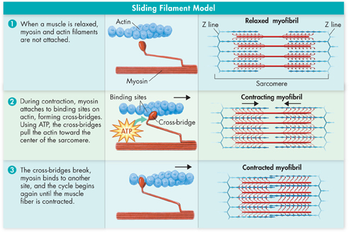
FIGURE 32–8 Sliding-Filament Model During muscle contraction, interaction between myosin filaments and actin filaments causes a muscle fiber to contract.
dddControl of Muscle Contraction Skeletal muscles are useful only if they contract in a controlled fashion. Remember that motor neurons connect the central nervous system to skeletal muscle cells. Impulses from these motor neurons control the contraction of muscle fibers.
A motor neuron and a skeletal muscle cell meet at a type of synapse known as a neuromuscular (noo roh MUS kyoo lur) junction. When a motor neuron is stimulated, its axon terminals release a neurotransmitter called acetylcholine (as ih til KOH leen). Acetylcholine (ACh) molecules diffuse across the synapse, producing an impulse (action potential) in the cell membrane of the muscle fiber. The impulse causes the release of calcium ions (Ca2+) within the fiber. These ions affect regulatory proteins that allow myosin cross-bridges to bind to actin.
A muscle cell contracts until the release of ACh stops and an enzyme produced at the axon terminal destroys any remaining ACh. Then, the muscle cell pumps Ca2+ back into storage, the cross-bridges stop forming, and the contraction ends.
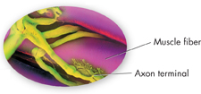
FIGURE 32–9 Neuromuscular Junction (SEM 825X)
MYSTERY CLUE

Children with rickets may suffer from muscle spasms. What might they be lacking that could cause uncontrolled muscle movements?
What is the difference between a strong contraction and a weak contraction? When you lift something light, such as a sheet of paper, your brain stimulates only a few cells to contract. However, as you exert maximum effort, such as when lifting your book bag, almost all the muscle cells in your arm are stimulated to contract.

Table of Contents
- Formulas and Equations
- Applying Formulas and Equations
- Mean, Median, and Mode
- Estimation
- Using Measurements in Calculations
- Effects of Measurement Errors
- Accuracy
- Precision
- Comparing Accuracy and Precision
- Significant Figures
- Calculating With Significant Figures
- Scientific Notation
- Calculating With Scientific Notation
- Dimensional Analysis
- Applying Dimensional Analysis