


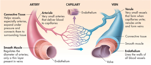
FIGURE 33–5 Structure of Blood Vessels The structure of blood vessel walls contributes to the vessels' functions.
dBlood Vessels
 What are three types of blood vessels?
Oxygen-rich blood leaving the left ventricle passes into the aorta. The aorta is the first of a series of vessels that carries blood through the systemic circulation and back to the heart. As blood flows through the circulatory system, it moves through three types of blood vessels—arteries, capillaries, and veins.
Arteries Arteries are large vessels that carry blood from the heart to the tissues of the body. Arteries are the superhighways of the circulatory system. Except for the pulmonary arteries, all arteries carry oxygen-rich blood. Arteries have thick elastic walls that help them withstand the powerful pressure produced when the heart contracts and pumps blood through them. Figure 33–5 describes the three layers of tissue found in artery walls—connective tissue, smooth muscle, and endothelium.
Capillaries The smallest blood vessels are the capillaries. Capillaries are the side streets and alleys of the circulatory system. Most capillaries are so narrow that blood cells pass through them in single file. Their extremely thin walls allow oxygen and nutrients to diffuse from blood into tissues, and carbon dioxide and other waste products to move from tissues into blood.
Veins After blood passes through the capillaries, it returns to the heart through veins. Blood often must flow against gravity through the large veins in your arms and legs. Many veins are located near and between skeletal muscles, as shown in Figure 33–6. When you move, the contracting skeletal muscles squeeze the veins, pushing blood toward the heart. Many veins contain valves. The valve that is farthest from the heart closes to ensure blood continues to flow in one direction.
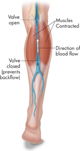
FIGURE 33–6 Blood Flow in Veins The contraction of skeletal muscles helps move blood in veins toward the heart. Draw Conclusions What role do valves play in large veins?
Table of Contents
- Formulas and Equations
- Applying Formulas and Equations
- Mean, Median, and Mode
- Estimation
- Using Measurements in Calculations
- Effects of Measurement Errors
- Accuracy
- Precision
- Comparing Accuracy and Precision
- Significant Figures
- Calculating With Significant Figures
- Scientific Notation
- Calculating With Scientific Notation
- Dimensional Analysis
- Applying Dimensional Analysis