


White Blood Cells White blood cells, or leukocytes (LOO koh syts), are the “army” of the circulatory system.  White blood cells guard against infection, fight parasites, and attack bacteria. The body can increase the number of active white blood cells dramatically during a “battle” with foreign invaders. In fact, a sudden increase in white blood cells is a sign that the body is fighting a serious infection. White blood cells are not confined to blood vessels. Many white blood cells can slip through capillary walls to attack foreign organisms.
Different types of white blood cells perform different protective functions. For example, macrophages engulf pathogens. Lymphocytes are involved in the immune response. B lymphocytes produce antibodies that fight infection and provide immunity. T lymphocytes help fight tumors and viruses. You will learn more about lymphocytes and other white blood cells in Chapter 35.
In a healthy person, white blood cells are outnumbered by red blood cells by almost 1000 to 1. Like red blood cells, white blood cells are produced from stem cells in bone marrow. Unlike red blood cells, however, white blood cells keep their nuclei and can live for years.
Platelets Blood loss can be life-threatening. Fortunately, a minor cut or scrape may bleed for a bit, but then the bleeding stops. Why? Because blood clots. Blood clotting is made possible by plasma proteins and cell fragments called platelets. The cytoplasm of certain bone marrow cells divides into thousands of small fragments. The fragments, each enclosed in a cell membrane, break off and enter the blood as platelets.
When platelets come in contact with the edges of a broken blood vessel, their surface becomes sticky, and they cluster around the wound. These platelets release proteins called clotting factors that start a series of reactions. Figure 33–9 summarizes one part of the clotting process.
 In Your Notebook Make a flowchart that describes the blood-clotting process.
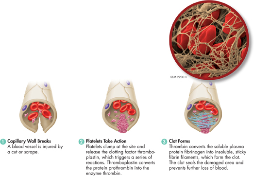
FIGURE 33–9 How Blood Clots Form This figure shows one chain reaction in the formation of a clot. When the clot is formed, strands of fibrin form a net that prevents blood from leaving the damaged vessel. Use Analogies How is a blood clot like a screened porch?
ddTable of Contents
- Formulas and Equations
- Applying Formulas and Equations
- Mean, Median, and Mode
- Estimation
- Using Measurements in Calculations
- Effects of Measurement Errors
- Accuracy
- Precision
- Comparing Accuracy and Precision
- Significant Figures
- Calculating With Significant Figures
- Scientific Notation
- Calculating With Scientific Notation
- Dimensional Analysis
- Applying Dimensional Analysis