


Hearing and Balance
 How do the ears and brain process sounds and maintain balance?
The human ear has two sensory functions, one of which, of course, is hearing. The other function is detecting positional changes associated with movement. Mechanoreceptors found in parts of the ear transmit impulses to the brain. The brain translates the impulses into sound and information about balance.
Hearing Sound is nothing more than vibrations moving through the air around us. The ears are the sensory organs that can distinguish both the pitch and loudness of those vibrations. The structure of the ear is shown in Figure 31–13.
Vibrations enter the ear through the auditory canal and cause the tympanum (TIM puh num), or eardrum, to vibrate. Three tiny bones, commonly called the hammer, anvil, and stirrup, transmit these vibrations to a membrane called the oval window. Vibrations there create pressure waves in the fluid-filled cochlea (KAHK lee uh) of the inner ear. The cochlea is lined with tiny hair cells that are pushed back and forth by these pressure waves. In response, the hair cells send nerve impulses to the brain, which processes them as sounds.
Balance Your ears contain structures that help your central nervous system maintain your balance, or equilibrium. Within the inner ear just above the cochlea are three tiny canals. They are called semicircular canals because each forms a half circle. The semicircular canals and the two tiny sacs located behind them monitor the position of your body, especially your head, in relation to gravity.
The semicircular canals and the sacs are filled with fluid and lined with hair cells. As the head changes position, the fluid in the canals also changes position. This causes the hair on the hair cells to bend. This action, in turn, sends impulses to the brain that enable it to determine body motion and position.
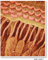
FIGURE 31–13 The Ear The diagram shows the structures in the ear that transmit sound. The SEM shows hair cells in the inner ear. The motion of these sensitive hair cells produces nerve impulses that travel to the brain through the cochlear nerve. Predict How would frequent exposure to loud noises that damage hair cells affect a person's threshold for detecting sound?
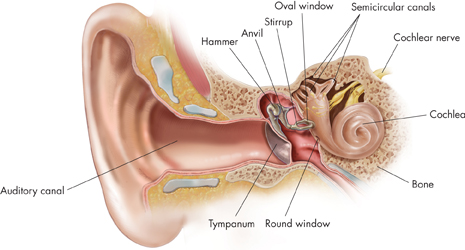
Table of Contents
- Formulas and Equations
- Applying Formulas and Equations
- Mean, Median, and Mode
- Estimation
- Using Measurements in Calculations
- Effects of Measurement Errors
- Accuracy
- Precision
- Comparing Accuracy and Precision
- Significant Figures
- Calculating With Significant Figures
- Scientific Notation
- Calculating With Scientific Notation
- Dimensional Analysis
- Applying Dimensional Analysis