


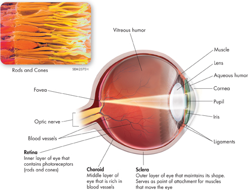
FIGURE 31–14 The Eye The eye is a complicated sense organ. The sclera, choroid, and retina are three layers of tissues that form the inner wall of the eyeball. Interpret Graphics What is the function of the sclera?
ddVision
 How do the eyes and brain produce vision?
The world around us is bathed in light, and the sense organs we use to detect that light are the eyes. Vision occurs when photoreceptors in the eyes transmit impulses to the brain, which translates these impulses into images.
Structures of the Eye The structures of the eye are shown in Figure 31–14. Light enters the eye through the cornea, a tough transparent layer of cells. The cornea helps to focus the light, which then passes through a chamber filled with a fluid called aqueous (AY kwee us) humor. At the back of the chamber is a disk-shaped structure called the iris. The iris is the colored part of the eye. In the middle of the iris is a small opening called the pupil. Tiny muscles in the iris adjust the size of the pupil to regulate the amount of light that enters the eye. In dim light, the pupil becomes larger and more light enters the eye. In bright light, the pupil becomes smaller and less light enters the eye.
Just behind the iris is the lens. Small muscles attached to the lens change its shape, helping to adjust the eyes' focus to see near or distant objects clearly. Behind the lens is a large chamber filled with a transparent, jellylike fluid called vitreous (VIH tree us) humor.

Table of Contents
- Formulas and Equations
- Applying Formulas and Equations
- Mean, Median, and Mode
- Estimation
- Using Measurements in Calculations
- Effects of Measurement Errors
- Accuracy
- Precision
- Comparing Accuracy and Precision
- Significant Figures
- Calculating With Significant Figures
- Scientific Notation
- Calculating With Scientific Notation
- Dimensional Analysis
- Applying Dimensional Analysis