


VISUAL SUMMARY
HOW SCIENTISTS MANIPULATE DNA
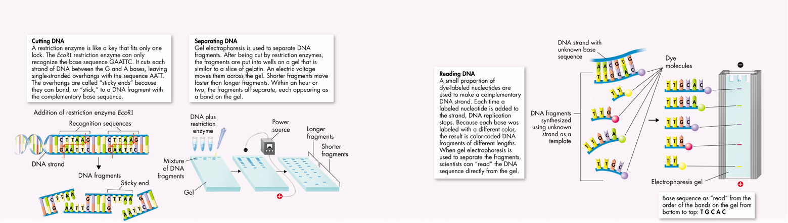
FIGURE 14–10 By using tools that cut, separate, and replicate DNA, scientists can read the base sequences in DNA from any cell. Knowing the sequence of an organism's DNA allows us to study specific genes.
Separating DNA Once DNA has been cut by restriction enzymes, scientists can use a technique known as gel electrophoresis to separate and analyze the differently sized fragments. Figure 14–10 illustrates this simple, yet effective, method. A mixture of DNA fragments is placed at one end of a porous gel. When an electric voltage is applied to the gel, DNA molecules—which are negatively charged—move toward the positive end of the gel. The smaller the DNA fragment, the faster and farther it moves. The result is a pattern of bands based on fragment size. Specific stains that bind to DNA make these bands visible. Researchers can then remove individual restriction fragments from the gel and study them further.
Reading DNA After the DNA fragments have been separated, researchers use a clever chemical “trick” to read, or sequence, them. The single-stranded DNA fragments are placed in a test tube containing DNA polymerase—the enzyme that copies DNA—along with the four nucleotide bases, A, T, G, and C. As the enzyme goes to work, it uses the unknown strand as a template to make one new DNA strand after another. The tricky part is that researchers also add a small number of bases that have a chemical dye attached. Each time a dye-labeled base is added to a new DNA strand, the synthesis of that strand stops. When DNA synthesis is completed, the result is a series of color-coded DNA fragments of different lengths. Researchers can then separate these fragments, often by gel electrophoresis. The order of colored bands on the gel tells the exact sequence of bases in the DNA. The entire process can be automated and controlled by computers, so that DNA sequencing machines can read thousands of bases in a matter of seconds.
Table of Contents
- Formulas and Equations
- Applying Formulas and Equations
- Mean, Median, and Mode
- Estimation
- Using Measurements in Calculations
- Effects of Measurement Errors
- Accuracy
- Precision
- Comparing Accuracy and Precision
- Significant Figures
- Calculating With Significant Figures
- Scientific Notation
- Calculating With Scientific Notation
- Dimensional Analysis
- Applying Dimensional Analysis